
Introduction
Uremic tumoral calcinosis (UTC) is an uncommon form of extraosseous calcification associated with advanced chronic kidney disease (CKD).1 It is characterized by the deposition of calcium phosphate crystals in periarticular soft tissues, typically around large joints.2 Its development has been linked to persistent alterations in mineral-bone metabolism, mainly sustained hyperphosphatemia, elevated calcium-phosphorus product, and secondary hyperparathyroidism.3 Although most reported cases involve the shoulders, elbows, or hips, distal phalangeal involvement is unusual and poorly documented in the literature.1,4 The relevance of this case lies not only in the atypical location but also in that the diagnosis is supported by a nearly 4-year longitudinal biochemical follow-up, allowing precise reconstruction of the metabolic substrate that favored ectopic calcification.5
Case report
A 47 year old female patient with end-stage CKD due to autosomal dominant polycystic kidney disease, on renal replacement therapy with hemodialysis twice weekly since May 2022, is presented. During evaluation in the context of nephrology follow-up, bilateral firm, non-tender swelling was incidentally identified in the distal phalanges of the first, second, and third fingers of the right hand, as well as the third finger of the left hand, with overlying desquamation and erythema (Fig. 1). These findings prompted a targeted workup.
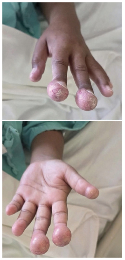
Figure 1. Clinical photographs of the patient showing swelling in the distal phalanges of the first, second, and third fingers of the right hand, with overlying trophic skin changes.
In the context of advanced CKD on renal replacement therapy, a mineral-bone metabolism disorder was proposed as the pathophysiological hypothesis. The immunological profile was negative, ruling out autoimmune or rheumatological etiology.
Admission laboratory studies showed intact parathyroid hormone (iPTH) of 1086.5 pg/mL, confirmed in duplicate – more than 16 times the upper normal limit – phosphorus of 6.8 mg/dL, alkaline phosphatase (ALP) of 347 U/L, and serum calcium of 9.7 mg/dL. Hand radiographs (March 12, 2026) revealed dense, periarticular, bilateral nodular calcifications in the soft tissues of the affected distal phalanges, compatible with UTC (Fig. 2).
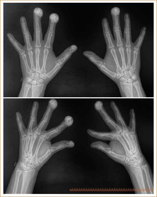
Figure 2. Hand radiographs of the patient (March 12, 2026). Dense nodular calcifications are observed in the periarticular soft tissues of the phalanges of the first, second, and third fingers of the right hand, and the third finger of the left hand, compatible with uremic tumoral calcinosis.
To define the metabolic substrate of this finding, a descriptive retrospective analysis of 47 months of biochemical follow-up was performed, from May 2022 to March 2026. Chronic hyperphosphatemia was observed with a mean of 5.96 ± 1.89 mg/dL and elevation in 75.4% of 69 measurements. ALP showed a mean of 252.9 ± 49.9 U/L, with persistent elevation in 100% of 29 measurements. Serum calcium remained within normal range, with a mean of 9.03 ± 0.43 mg/dL, with no elevated measurements in 70 determinations. Creatinine showed a mean of 9.49 ± 2.75 mg/dL, elevated in 100% of 77 measurements, consistent with persistent severe renal dysfunction. The statistical summary is presented in table 1.
Table 1. Descriptive statistical analysis of biochemical parameters during 47 months of follow-up (May 2022 to March 2026)
| Parameter | n | Mean ± SD | Range | Percentage elevated |
|---|---|---|---|---|
| Phosphorus (mg/dL) | 69 | 5.96 ± 1.89 | 1.9-10.5 | 75.4 |
| Urea (mg/dL) | 78 | 99.39 ± 60.54 | 16.0-235.0 | 78.2 |
| ALP (U/L) | 29 | 252.9 ± 49.9 | 124-372 | 100 |
| Calcium (mg/dL) | 70 | 9.03 ± 0.43 | 7.0-10.2 | 0 |
| Creatinine (mg/dL) | 77 | 9.49 ± 2.75 | 4.33-17.2 | 100 |
|
Reference values: Phosphorus 2.5-4.5 mg/dL; Urea 17-43 mg/dL; ALP 25-100 U/L; Calcium 8.5-10.5 mg/dL; Creatinine 0.6-1.1 mg/dL.ALP: alkaline phosphatase; SD: standard deviation. |
||||
Discussion
The main contribution of this case is the documentation of an atypical presentation of UTC in distal phalanges, supported by objective longitudinal biochemical evidence.5 Clinically, this is not merely an unusual location, but a visible manifestation of accumulated chronic metabolic exposure. The patient presented an approximately 4-year course with persistent impairment of phosphocalcic metabolism: hyperphosphatemia present in three out of four measurements and constant ALP elevation, an indirect marker of active bone remodeling.3 This was compounded by severe secondary hyperparathyroidism with PTH exceeding 1000 pg/mL, reinforcing the biological environment favoring ectopic deposition of calcium phosphate crystals.6,7
Another relevant aspect is the value of longitudinal follow-up. In many case reports, interpretation relies on isolated measurements; in contrast, this case made it possible to demonstrate the pattern, persistence, and magnitude of exposure to the main risk factors.5 Phosphorus was not only elevated at a single point in time, but in 75.4% of 69 measurements; ALP was not only high at admission, but in 100% of 29 determinations; and iPTH at admission evidenced an extreme degree of secondary hyperparathyroidism. This temporal continuity strengthens the causal relationship between the mineral-bone disorder and the development of calcinosis.3
The distal location in the fingers deserves particular emphasis. UTC is typically described in large joints;2,4 therefore, manifestations in the phalanges may go unnoticed, be misinterpreted as dermatological, rheumatological, or infectious pathology, and delay diagnosis.1 In this case, negative autoantibodies and the bilateral periarticular nodular radiographic pattern helped exclude differential diagnoses and consolidate the correct etiological diagnosis.
It should be noted that the diagnosis was established using clinical, biochemical, and imaging criteria, without histopathological confirmation. While biopsy represents the definitive diagnostic standard, the literature recognizes that in the setting of advanced CKD with a characteristic metabolic profile and compatible radiological pattern, clinical-radiological diagnosis is acceptable and widely practiced.2,6 The absence of biopsy constitutes a limitation of this report that must be acknowledged; however, the concordance between the clinical context, the documented longitudinal metabolic substrate, and the radiographic findings provides sufficient diagnostic validity.
From a clinical practice standpoint, this case underscores that serial mineral-bone metabolism monitoring should not be considered an administrative requirement, but rather a tool for preventing extraskeletal complications.3,8 The presence of sevelamer in the therapeutic regimen and, despite this, persistent chronic hyperphosphatemia, suggests the need to reassess adherence, dosing, dietary phosphorus load, and dialytic adequacy.9 In patients with end-stage CKD on dialysis, continuous monitoring of phosphorus, calcium, PTH, and ALP can identify risk trajectories before irreversible calcifications develop.3,8
Conclusion
UTC should be considered in all patients with advanced CKD presenting with periarticular masses or swellings, even when the location is unusual.1,2 The main relevance of this case lies in two elements: the atypical presentation in distal phalanges, and the objective demonstration – through 47 months of follow-up – of chronic hyperphosphatemia, persistently elevated ALP, and severe secondary hyperparathyroidism with maintained normocalcemia. Together, these findings show that cumulative metabolic exposure may be more determinant than an isolated measurement in explaining ectopic calcification.10,11 This case supports the need for strict longitudinal monitoring of mineral-bone metabolism in hemodialysis patients.3,8
Funding
The authors declare that this work was carried out with the authors’ own resources.
Conflicts of interest
The authors declare that they have no conflicts of interest.
Acknowledgments
The authors thank the patient for her willingness to allow the publication of this case report.
Ethical considerations
Protection of human subjects and animals. The authors declare that no experiments on humans or animals were performed for this research.
Confidentiality, informed consent, and ethical approval. The authors have followed their institution’s confidentiality protocols, obtained informed consent from all patients, and secured approval from the Ethics Committee. SAGER guidelines have been followed as applicable to the nature of the study.
Declaration on the use of artificial intelligence. The authors declare that no generative artificial intelligence was used in the writing or creation of the content of this manuscript.
